SLEEP PARALYSIS
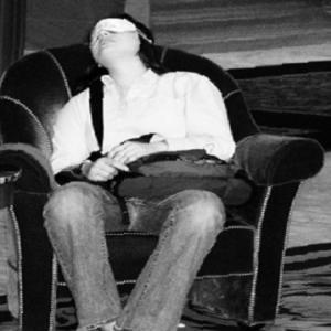
Sleep paralysis is characterized by the sensation of being unable to move while awakening from sleep. Sleep paralysis is described as a transitional state that occurs when a person experiences a temporary inability to react, move, or speak while asleep, falling asleep, or on awakening from sleep.
Symptoms of Sleep Paralysis
Unfortunately, sleep paralysis can be accompanied by frightening hallucinations during sleep whereby, due to the paralysis and physical experiences (which could be a forceful current running through the upper body), the person is left physically unable to react. These hallucinations can be very scary, and often involve a supernatural creature or other person taunting or terrifying the individual, together with difficulty breathing and/or a feeling of pressure on one’s chest. Another quite common type of hallucination involves either supernatural or human intruders lurking outside the person’s window or entering their bedroom, leaving the sleeper with feelings of fear and dread.1
During sleep paralysis, the individual is completely aware of the surroundings. The person often has the desire to move but is unable to. It can be frightening to the person.
What Causes Sleep Paralysis?
One explanation of sleep paralysis is that it’s caused by disrupted REM sleep: REM sleep typically induces total muscle atonia which prevents sleepers from acting out their dreams. Sleep deprivation and genetics are the major causes of sleep paralysis, and this condition has also been linked to disorder s such as migraines, narcolepsy, obstructive sleep apnea, and anxiety disorders. It is associated with excessive daytime sleepiness and being sleep deprived. When a person sleeps in a fixed supine position, it increases the likelihood of them experiencing sleep paralysis.In addition,it is related to Rapid eye movement (REM )atonia, which is the paralysis that occurs as a natural part of REM sleep. Sleep paralysis is also one of the symptoms of narcolepsy.
s such as migraines, narcolepsy, obstructive sleep apnea, and anxiety disorders. It is associated with excessive daytime sleepiness and being sleep deprived. When a person sleeps in a fixed supine position, it increases the likelihood of them experiencing sleep paralysis.In addition,it is related to Rapid eye movement (REM )atonia, which is the paralysis that occurs as a natural part of REM sleep. Sleep paralysis is also one of the symptoms of narcolepsy.When Does Sleep Paralysis Occur?
These events often occur when a person is either falling asleep or awakening from sleep.
If it occurs when going to sleep, the person will remain alert while the body prepares for REM sleep. This condition is known as predormital or hypnagogic sleep paralysis. If it occurs when the person is waking up, the person becomes alert prior to the REM cycle being completed. This condition is known as post-dormital or hypnopompic paralysis. The events can last anywhere from a few seconds to a few minutes, with rare cases lasting for hours, where the person could well experience panic symptoms.3
Sleep Paralysis Is Not Complete Paralysis
Due to the correlation of the paralysis with REM sleep, this type of atonia is not complete. The use of EOG traces clearly show that eye movement is still possible during these episodes; however, the person who is experiencing the events is not able to speak.
Comments
Post a Comment